

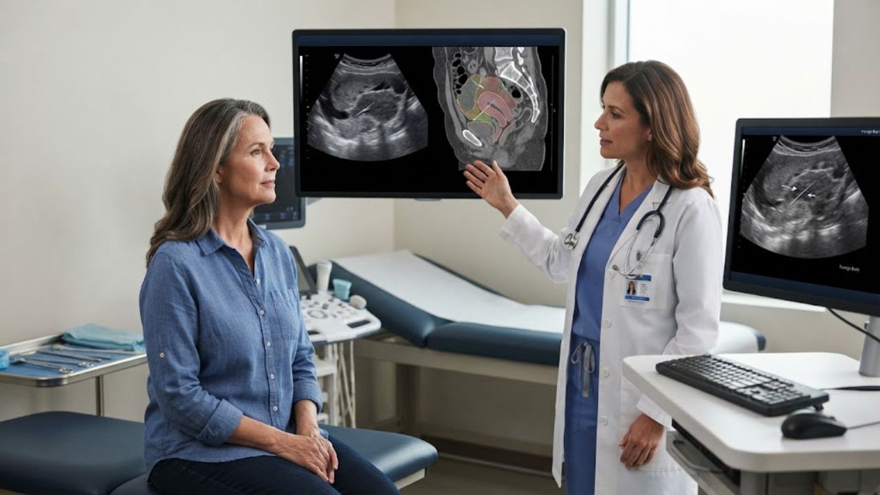
A 55-year-old woman in Chengdu, China, had been passing blood in her urine on and off for six months without pain and had never had it looked at. Two days before she finally came in, straining brought on heavy bleeding along with urinary frequency and urgency. An ultrasound found the back wall of her bladder irregularly thickened over an area measuring about 5.3 by 1.9 by 5.5 centimeters, with dense blood flow signals running through it. The reading pointed toward a malignant tumor.
It was a fish bone. Roughly 3 centimeters of it, lodged at the pelvic floor with one end in the rectal wall, the other pushed through the bladder muscle, and one side embedded in the outer layer of the uterus. The case report was published in Frontiers in Oncology.
Six Months of Blood and a Mass That Behaved Like a Tumor
Painless blood in the urine is a classic cancer warning sign, which is why the initial workup went where it did. Urologists performed a transurethral resection biopsy and found a broad-based, cauliflower-shaped growth about 5 by 2 centimeters on the back wall of the bladder, richly supplied with vessels and bleeding.
The pathology came back as chronic inflammation of the bladder lining with inflammatory granulation tissue underneath, not malignancy. Her white cell count, C-reactive protein, liver and kidney function and tumor markers were all unremarkable. She had no surgical history that might explain a retained object. Cervical cytology was negative. Three days after the biopsy, the bleeding returned.
At that point the case stopped fitting either explanation neatly, which is what pushed the team to keep imaging.
The Clue Was a Thin Line That Appeared on Every Scan
Contrast CT showed the thickened bladder wall blending into the uterine isthmus, with an unexplained linear dense shadow between them. MRI showed the same structure as a thin dark line that did not take up contrast, which the radiologists read as either post-biopsy change or a foreign body. Transvaginal ultrasound then showed a hypoechoic mass roughly 4.5 by 2.3 by 1.9 centimeters filling the space between uterus and bladder, with a bright linear band running continuously from the uterine isthmus into the bladder wall.
Bright on CT, dark on MRI, bright on ultrasound. That combination describes something hard, dense and acoustically resistant: bone, a stone or metal. The line was also continuous and regular rather than scattered, which argued against tumor calcification and for an object that did not belong there.
The authors were blunt about the trap. The initial imaging concentrated on the inside of the bladder, where a bladder tumor would be, and gave limited views of the structures outside it. The clinical picture had already suggested cancer, and the workup followed that assumption.
How a Swallowed Bone Reaches the Pelvis
Most swallowed fish bones lodge in the throat or esophagus and announce themselves immediately. Some reach the stomach, where acid softens smaller fragments enough for them to break down and pass.
Larger fragments keep their sharp edges. When one moves into the lower digestive tract, peristalsis cannot break it up and instead drives it against the bowel wall. It can ulcerate through, causing peritonitis, abscess or an inflammatory pseudotumor. Migrating bones have been documented reaching the liver and pancreas, and a previous report described a liver abscess from a fish bone that was initially read as metastatic cancer.
This patient never had the usual signs. No throat symptoms, no abdominal tenderness, no rebound pain. Over months the bone irritated the surrounding tissue into granulation and scar that wrapped around it, producing exactly the vascular, ill-defined mass that imaging interpreted as tumor. A fish bone has previously been found embedded in the bladder wall, and one earlier report described a bone that perforated the rectosigmoid colon and migrated to the bladderduring treatment, but the authors describe a bone spanning three organs at once as exceptionally rare.
Cutting the Bone Instead of Pulling It Out
Surgery was done laparoscopically with endoscopic assistance. The team found a hard mass about 5 by 3 centimeters at the pelvic peritoneal reflection, inflamed and stuck fast to the rectal wall, bladder and uterine isthmus, with the normal anatomy obscured.
Pulling a bone that crosses three organs would risk tearing all three. Instead the surgeons freed the adhesions in stages, cut the bone, and removed it in segments through combined endoscopic access. Final pathology confirmed inflammatory change with no malignant cells.
An ultrasound a week later showed only a scar-like area between uterus and bladder. At one month her bleeding, urinary frequency, urgency and painful urination had resolved, and repeat CT of the abdomen and pelvis was clear. European guidance on foreign body removal recommends choosing among endoscopic routes based on symptoms, overall condition and imaging, with open surgery reserved for emergencies or incomplete retrieval. A recent review of fish bone migration covers the diagnostic difficulty in more depth.
This is one patient. It does not mean unexplained blood in the urine is usually a foreign body, and painless hematuria still warrants prompt evaluation.
Key Questions Answered
What was actually wrong with the patient?
A fish bone about 3 centimeters long had perforated her rectum, the outer layer of her uterus and her bladder wall, creating an inflammatory mass.
Why was it mistaken for cancer?
Painless blood in the urine plus a vascular, ill-defined bladder wall mass fits bladder cancer. Scar and granulation tissue around the bone mimicked a tumor on imaging.
What finally revealed it?
A thin line that was dense on CT, dark on MRI and bright on ultrasound, running continuously between the uterus and the bladder wall.
How was it removed?
Surgeons separated the adhesions laparoscopically, cut the bone and removed it in pieces using combined endoscopic access rather than pulling it out whole.
Did she recover?
Yes. At one month her symptoms had resolved and repeat CT of the abdomen and pelvis showed no abnormalities.
Does this mean blood in the urine is often harmless?
No. Painless hematuria is a recognized cancer warning sign and needs prompt medical evaluation.


