

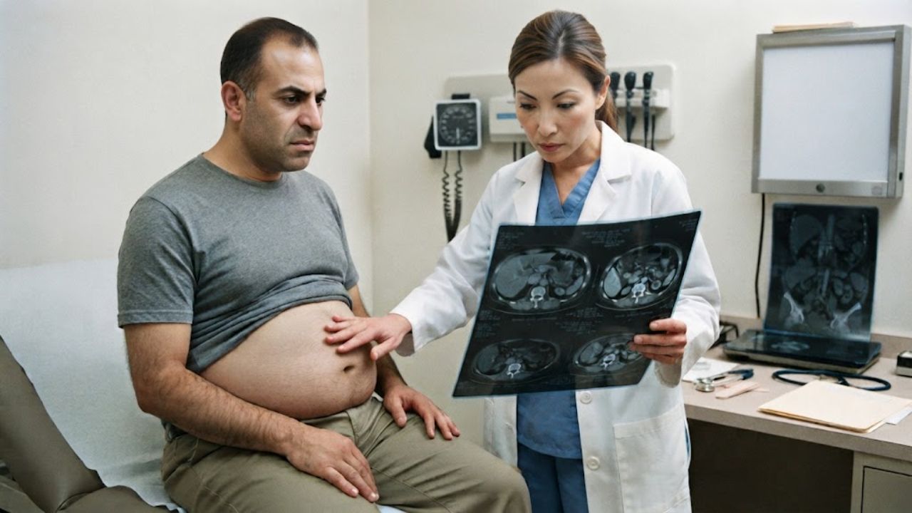
A 53-year-old man was referred to a surgical clinic for what looked like a straightforward problem: a hernia above his navel and months of abdominal pain. He had also lost 10 kilograms, roughly 22 pounds, and had been chronically constipated.
His scans complicated things. Ultrasound showed fluid in the abdomen and thickened omentum. A CT scan added an irregular liver outline, which in a man with type 2 diabetes could plausibly mean early fatty liver disease. It would have been an easy call to make and the wrong one.
When surgeons went in to look, the fluid in his abdomen was not fluid in the ordinary sense. It was mucin, thick and jelly-like, with nodules scattered across the peritoneal surfaces. The case was published February 27 in Cureus by clinicians at the University of Sharjah and Kuwait Hospital in the United Arab Emirates.
The Disease That Fills the Abdomen with Jelly
Pseudomyxoma peritonei occurs when mucin-producing tumor cells spread across the lining of the abdominal cavity and keep secreting. The mucin accumulates. Pressure rises. Organs get compressed.
The nickname doctors use for it, and the one the authors put in print, is jelly belly.
It is genuinely rare, with an estimated incidence of one to two cases per million people per year. The appendix is the most common source. In this patient, the appendiceal tip looked abnormal and nodular during laparoscopy, and pathology confirmed a low-grade appendiceal mucinous neoplasm, graded I under World Health Organization criteria. Biopsies from the omentum and peritoneal nodules showed low-grade mucinous neoplasia.
Mean age at onset is around 53, and the condition is slightly more common in women. In men, it usually traces back to a ruptured mucinous lesion of the appendix.
Grading is not a technicality here. An international expert panel established a standardized classification for the disease and its associated appendiceal tumors, separating low-grade from high-grade mucinous carcinoma peritonei. The two forms carry substantially different outlooks, and the distinction drives how aggressively patients are treated. This man’s disease fell in the low-grade category.
The Hernia Was a Clue, Not a Coincidence
The supraumbilical hernia that brought him to the clinic in the first place is the detail the authors highlight.
New-onset hernias are a recognized but infrequent presentation of pseudomyxoma peritonei, and at least one earlier report described a recurrent incisional hernia caused by the condition. The mechanism is simple mechanics. Mucin accumulating inside a closed cavity raises intra-abdominal pressure steadily over months or years, and the abdominal wall eventually gives at its weakest point. His CT also found four small paraumbilical hernias, each about 5 millimeters.
That is why the diagnosis is so often missed or delayed. Distension, changed bowel habits, appetite loss, and weight loss are nonspecific, and when they arrive with abdominal fluid, chronic liver disease is a far more common explanation. Many cases are found incidentally during surgery for something else entirely.
The Signs Radiologists Look For
Three imaging findings pointed away from liver disease and toward the peritoneum.
Omental caking, a thickening of the fatty apron that drapes the abdominal organs, showed on ultrasound and again on MRI with restricted diffusion. Septated peritoneal fluid collections appeared on MRI. Hepatic scalloping, which the authors call the most characteristic radiological sign of the condition, showed indentations pressed into the surface of the liver by the thick mucin.
Contrast-enhanced CT can distinguish mucinous material from ordinary fluid because the two differ in attenuation, with mucin showing low-attenuation areas mixed with denser solid tumor elements.
His tumor markers were partly informative. CA 19-9 and alpha-fetoprotein were normal. Carcinoembryonic antigen was elevated at 10.2 to 18.0 nanograms per milliliter against a reference ceiling below 3.8, roughly three to five times the upper limit. The authors note that low grade disease is generally associated with more modest CEA elevations than high-grade disease.
Even so, imaging and blood work were not enough. The differential still included peritoneal carcinomatosis, tuberculous peritonitis, and peritoneal mesothelioma. Only tissue settled it.
What Happens Next for Patients Like Him
Untreated, pseudomyxoma peritonei progresses and can be fatal through bowel obstruction, malnutrition, and infection.
The standard of care is cytoreductive surgery combined with hyperthermic intraperitoneal chemotherapy, in which heated chemotherapy is circulated inside the abdominal cavity after the visible tumor is removed. A 2025 multisociety consensus statement formally evaluated the evidence behind that combination. Studies cited by the authors report five year survival rates ranging from 62 to 80 percent following complete cytoreduction plus the heated chemotherapy, with low-grade disease carrying the better outlook.
This patient recovered without incident from his diagnostic laparoscopy, appendectomy and biopsies, and was referred to a specialized oncology center for that combined treatment. He returned weeks later with left-sided kidney colic, and repeat imaging still showed ascites, omental thickening and hepatic scalloping.
The authors emphasize “the importance of maintaining a high index of suspicion” in patients with unexplained abdominal fluid, increasing girth, and abdominal wall hernias. This is one patient’s experience and does not establish how often hernias signal the condition. Anyone with a persistently swelling abdomen, unexplained weight loss, or a new hernia should be evaluated rather than assume a familiar explanation fits.
Key Questions Answered
What is pseudomyxoma peritonei? A rare condition in which mucin producing tumor cells spread across the lining of the abdominal cavity and continue secreting thick mucinous material. The accumulation enlarges the abdomen and compresses organs. Doctors nickname it jelly belly.
How rare is it? Estimated at one to two cases per million people per year. Mean age at onset is around 53, and it occurs slightly more often in women than men.
Where does it come from? Most often from a low grade mucinous neoplasm of the appendix. Less commonly from the ovary, colon, urachus or other sites. In women the ovaries are frequently involved secondarily to an appendiceal primary.
Why is a hernia significant here? Mucin building up inside the abdomen raises internal pressure over time, which can push the abdominal wall open at a weak point. New onset hernias are an infrequent but recognized presentation of the condition.
How is it diagnosed? Imaging findings including omental caking, septated fluid collections and hepatic scalloping raise suspicion, but definitive diagnosis requires direct visualization and tissue biopsy. Tumor markers, particularly CEA, support diagnosis and follow up.
What is the treatment and outlook? Cytoreductive surgery combined with heated intraperitoneal chemotherapy is the standard approach. Cited studies report five year survival of 62 to 80 percent after complete cytoreduction, with low grade disease carrying the better prognosis.


