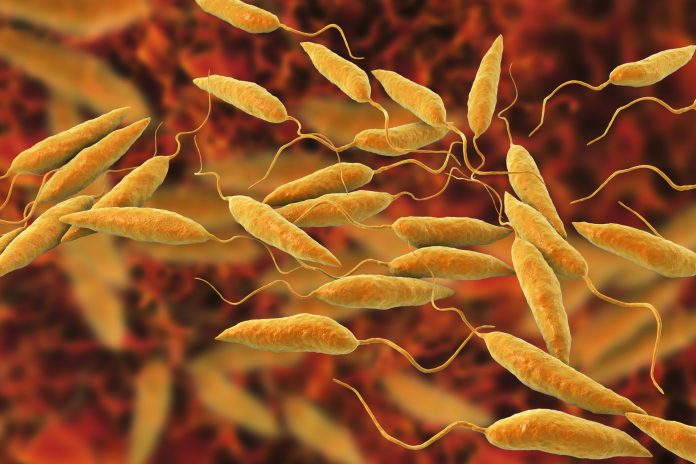
Infectious agent
Leishmaniasis is caused by obligate intracellular protozoan parasites; >20 Leishmania species cause leishmaniasis. Leishmaniasis has different forms, including visceral leishmaniasis (VL), cutaneous leishmaniasis (CL), and mucosal leishmaniasis (ML). The presenting form varies by Leishmania species. For example, Leishmania infantum and Leishmania donovani are the most common causes of VL, while ML is most often associated with Viannia species.
Transmission
Leishmania parasites are most often transmitted through the bites of infected female phlebotomine sand flies. Transmission risk is greatest from dusk to dawn because sand flies typically feed (i.e., bite) at night and during twilight hours. Vector activity might easily be overlooked because sand flies are small and silent, and their bites can go unnoticed. Travelers with potentially increased risk for leishmaniasis include adventure travelers, bird watchers, construction workers, ecotourists, military personnel, missionaries, Peace Corps volunteers, and people doing research or humanitarian work outdoors at night or twilight. However, even short-term travelers in leishmaniasis-endemic areas have developed leishmaniasis. Immigrants and refugees from endemic areas also might present with leishmaniasis. A less common mode of transmission for both CL and VL is accidental occupational (laboratory) exposures. For VL, uncommon modes include congenital transmission, blood transfusions, organ transplantation, and sharing of contaminated needles.
Epidemiology
In 2021, leishmaniasis was reported to be endemic in 99 countries. An estimated 600,000 to 1,000,000 new cases of CL and 50,000 to 90,000 new cases of VL occur annually. The ecologic settings for leishmaniasis transmission range from rainforests to arid regions.
In the Eastern Hemisphere, CL is found in Africa, particularly the tropical region and North Africa; Asia, particularly Central and Southwest Asia; southern Europe, including southern France, Greece, Italy, Portugal, Spain, and the Mediterranean islands; and some countries of the Middle East. Leishmaniasis from these areas is often referred to as “Old World” leishmaniasis. In the Western Hemisphere, CL is found in parts of Mexico, all countries of Central America, and most of South America except Chile and Uruguay. Endemic transmission in the United States has been identified in Texas, especially among people living in areas bordering northeastern Mexico, and possibly in Arizona and Oklahoma. Leishmaniasis from these areas is often referred to as “New World” leishmaniasis. Endemic transmission of CL is not found in Canada. Visit the World Health Organization (WHO) cutaneous leishmaniasis endemicity map for more information.
GeoSentinel Surveillance from 1997–2017 indicated that among patients with laboratory confirmed CL examined at specialized travel or tropical medicine clinics on 6 continents, including North America, common source countries for travel-associated CL were in the Amazon Basin (Brazil, Bolivia, Colombia, Ecuador, and Peru); Costa Rica; El Salvador; and Israel. Among immigrants, common source countries were Afghanistan and Syria. Cases of CL in U.S. service personnel have reflected military activities (e.g., in Afghanistan and Iraq). CL is usually more common in rural than urban areas but is found in some peri-urban and urban areas (e.g., in Kabul, Afghanistan).
Although ML is uncommon, it has occurred in travelers and expatriates, including in people whose cases of CL were not treated or were treated inadequately. ML is most commonly associated with the Viannia subgenus found in Central and South America, particularly in infections acquired in Bolivia, Brazil, or Peru. However, ML has been documented on rare occasions with species of Leishmania found in various countries of the Eastern Hemisphere.
VL is usually more common in rural than urban areas, but it is found in some peri-urban areas (e.g., in northeastern Brazil). In the Eastern Hemisphere, VL is found in Africa, particularly East Africa; parts of Asia, particularly the Indian subcontinent and Central and Southwest Asia; southern Europe; and the Middle East. In the Western Hemisphere, most cases occur in Brazil; some cases occur in scattered foci elsewhere in Latin America. Overall, VL is found in focal areas of >70 countries. In 2021, 97% of reported VL cases were from 11 countries: Brazil, Eritrea, Ethiopia, India, Kenya, Nepal, Somalia, South Sudan, Sudan, Uganda, and Yemen. Visit the 2020 WHO visceral leishmaniasis endemicity map for more information.
The geographic distribution of VL cases evaluated in the United States and other countries reflects travel and immigration patterns. Although uncommon in most U.S. travelers, expatriates, and military personnel, VL has occurred in such short-term and long-term travelers returning from travel or deployment to endemic regions in European countries (e.g., France, Greece, Italy, Macedonia, and Spain) and the Middle East.
Clinical presentation
Clinical presentation depends on the form of leishmaniasis (i.e., CL, ML, or VL). Not all people with Leishmania infection develop symptoms.
Cutaneous leishmaniasis
CL can present with a broad variety of dermatologic manifestations ranging from small and localized skin lesions to large nodules or plaques covering multiple body surfaces. The clinical spectrum can mimic other skin conditions (e.g., leprosy, squamous cell cancer, fungal or other skin infections).
Lesions typically develop on exposed areas of the skin within several weeks after infection. However, lesions may first appear months or years later, often in the context of trauma (e.g., skin wounds, surgery). The lesions can change in appearance and size over time, typically progressing from small, erythematous papules or nodular plaques to open sores with raised borders and central craters (i.e., ulcers), which can be covered with crusts or scales. Lesions usually are painless but can be painful if superinfected with bacteria. Satellite lesions, regional lymphadenopathy, and nodular lymphangitis can occur. Even without treatment, most lesions eventually heal; they can last for months or years, however, and typically result in scarring.
Mucosal leishmaniasis
Some Leishmania species are a potential concern because parasites might spread from the skin to the mucosal surfaces of the nose or mouth and cause lesions in these areas. Most often, these are Leishmania Viannia species in Central and South America. ML frequently appears years after the original skin lesions have healed but may also occur along with the skin lesions or as the initial presentation of a subclinical cutaneous infection. The initial clinical manifestations typically involve the nose, with bleeding, chronic stuffiness, and inflamed mucosa or lesions; less often the mouth or larynx are involved, manifesting as a brassy cough or hoarseness. In advanced cases, ulcerative destruction of the mouth, nose, larynx, and pharynx (e.g., perforation of the nasal septum, laryngeal damage, or tracheal damage) can occur.
Visceral leishmaniasis
Among symptomatic people, the incubation period typically ranges from weeks to months. Illness onset can be abrupt or gradual. Stereotypical clinical manifestations of VL include fever, hepatosplenomegaly (especially splenomegaly), night sweats, and weight loss; lymphadenopathy can occur. Laboratory findings characteristic of VL include pancytopenia (anemia, leukopenia, thrombocytopenia), high total protein, low albumin, and hypergammaglobulinemia. If untreated, severe (advanced) cases of VL typically are fatal. Latent infection can clinically manifest years to decades after exposure in people who become immunocompromised through HIV infection, biologic immunomodulatory therapy, or immunosuppressive therapy.
Diagnosis
CDC can assist with the diagnostic evaluation of suspected leishmaniasis, including parasite species identification. For consultative services, contact CDC Parasitic Diseases Inquiries (404-718-4745; parasites@cdc.gov). CDC’s Parasitology Lab (404-718-4175; parasiteslab@cdc.gov) can provide helpful information regarding collection, storage, and shipping of samples for Leishmania diagnostic testing.
Cutaneous and mucosal leishmaniasis
Consider CL in people with chronic, non-healing skin lesions who have been in areas where leishmaniasis is found. Clinical signs and symptoms are not sufficiently specific to differentiate CL from other conditions. Obtain an explicit travel history, including, if possible, questioning fellow travelers about similar lesions. Obtain information about duration and progression of symptoms, whether the lesions are painful, prior treatment, and current medications (e.g., immunosuppressive agents); photographs are helpful to assess lesions over time. Conduct a careful physical examination, including evaluation of skin, lymph nodes, and mucosal surfaces.
Any patient with CL caused by a species with risk of causing ML, primarily related to the Viannia subgenus species (Leishmania (V.) braziliensis, Leishmania (V.) guyanensis, and Leishmania (V.) panamensis), but also rarely related to some additional New World and Old World species, should undergo a careful examination of mucosal surfaces, including the vocal cords and oronasal pharynx, along with biopsy of any abnormal appearing tissue, to evaluate for ML. This mucosal exam should occur regardless of other symptoms. Referral to a specialist able to conduct an endoscopic laryngeal examination is warranted if ML is suspected.
Laboratory confirmation of the diagnosis is achieved by detecting Leishmania parasites or DNA in infected tissue through light-microscopic examination of stained specimens, culture techniques, or molecular methods (e.g., polymerase chain reaction); conducting multiple tests on the specimen improves diagnostic yield. Because different Leishmania species have different management implications, species identification through molecular testing is important, particularly if >1 species is endemic to areas where the patient traveled. Serologic testing generally is not useful for CL because the assays are insensitive and cannot distinguish between active and past infection.
Visceral leishmaniasis
Consider VL in the differential diagnosis of people with a relevant travel history, even in the distant past, and a persistent, unexplained febrile illness, especially if accompanied by other suggestive manifestations (e.g., pancytopenia or splenomegaly). Hemophagocytic lymphohistiocytosis could be a complication and should prompt healthcare professionals to consider VL in patients with the appropriate travel history.
Laboratory confirmation of the diagnosis is achieved by detecting Leishmania parasites or DNA in infected blood, bone marrow, liver, or lymph nodes through light-microscopic examination of stained specimens, molecular methods, or tissue culture techniques. Serologic testing can provide supportive evidence for the diagnosis.
Treatment
Treatment of leishmaniasis can be complex; healthcare professionals can receive expert consultation from CDC staff about treatment approaches (see the “Diagnosis” section of this chapter for contact information).
Cutaneous and mucosal leishmaniasis
The primary goal of CL and ML treatment is to prevent morbidity. Decisions about whether and how to treat CL, including whether to use a systemic (oral or parenteral) medication rather than a local or topical approach, should be individualized. All cases of ML should be treated with systemic therapy. The response to a particular regimen can vary not only among Leishmania species but also for the same species in different geographic regions. Expert consultation is advised for treatment of any patient with CL, ML, or VL.
Some patients with CL may be candidates for local therapy depending on the risk of mucosal dissemination, patient characteristics, and the characteristics, number, and location of the lesion(s). Local therapy can include cryotherapy, heat therapy, topical paromomycin, or intralesional pentavalent antimonials. In the United States, topical paromomycin may be available through compounding pharmacies and its use is considered off-label. The oral agent miltefosine is approved by the U.S. Food and Drug Administration (FDA) to treat CL caused by 3 Western Hemisphere species of the Viannia subgenus: Leishmania (V.) braziliensis, L. (V.) guyanensis, and L. (V.) panamensis, as well as for ML caused by L. (V.) braziliensis in adults and adolescents ≥12 years old who weigh ≥30 kg and are not pregnant or breastfeeding during therapy or for 5 months after the last dose of miltefosine. Various parenteral options, including liposomal amphotericin B, are commercially available, although not FDA-approved to treat CL or ML. The pentavalent antimonial compound meglumine antimoniate (Glucantime) is not FDA-approved but can be acquired through an Investigational New Drug (IND) application. Glucantime can be administered intravenously, intramuscularly, or intralesionally; the route of administration should be individualized, and expert consultation is advised. Pentostam is no longer available, but sodium stibogluconate may be available in some countries outside of the United States.
Visceral leishmaniasis
All symptomatic patients with VL should be treated. Refer patients with VL to an infectious disease or tropical medicine specialist who can help direct care and provide individualized treatment. Risk for relapse and treatment failure is greater in immunocompromised VL patients compared with immunocompetent VL patients, particularly those living with HIV. Therefore, treatment of VL can differ between immunocompromised and immunocompetent VL patients. Relapse and treatment failure rarely might occur in immunocompetent patients.
Liposomal amphotericin B (AmBisome) is approved by the FDA to treat VL and is generally the drug of choice for U.S. patients. The oral agent miltefosine is approved by the FDA to treat VL in patients infected with L. donovani who are ≥12 years old, who weigh ≥30 kg, and who are not pregnant or breastfeeding during therapy or for 5 months after treatment. Pentavalent antimonials (e.g., meglumine antimoniate [Glucantime], sodium stibogluconate) are used in endemic areas, except for India, where developing resistance is a concern. See the “Cutaneous and Mucosal Leishmaniasis” section above for further information on the availability of pentavalent antimonials.
Prevention
No vaccines or drugs to prevent infection are available. Travelers can reduce the risk of CL by using personal protective measures to avoid sand fly bites (see Mosquitoes, Ticks, and Other Arthropods chapter). Advise travelers to avoid outdoor activities, especially from dusk to dawn when sand flies are the most active; wear protective clothing and apply insect repellent to exposed skin and under the edges of clothing (e.g., shirt sleeves, pant legs) according to the manufacturer’s instructions; and sleep in air-conditioned or well-screened areas. Spraying sleeping quarters with insecticide might provide some protection, and fans or ventilators might inhibit the movement of sand flies, which are weak fliers.
Sand flies are small (approximately 2–3 mm,


