

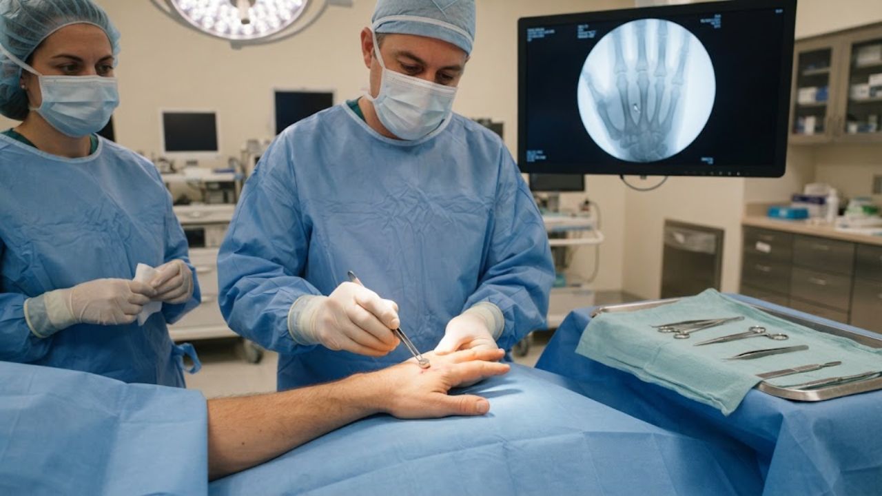
Metal fragments end up in hands and forearms in unglamorous ways: a metalworking accident, a surgical needle snapping mid-procedure, a displaced acupuncture instrument, a shot from a BB gun. Left in place they can produce chronic pain, infection, bone infection or reactive scarring, and digging them out carries its own risk of nicking a nerve or vessel.
Getting one out normally means an incision, a search through soft tissue, and a run of X-ray images while the surgeon hunts for something too small to feel. A team in Istanbul reported doing it a different way. Hold a sterilized magnet against the skin, find the spot where the pull is strongest, make a small cut, and let the fragment come to the magnet.
Across 15 consecutive patients treated over two years, all 15 fragments came out. The average operation took 3.13 minutes. The average number of fluoroscopy images was 0.88 per case, 13 across the entire series. The report was published in Frontiers in Surgery.
Finding the Spot by Feel
The instruments were not exotic. The main one was a commercially available neodymium-iron-boron disc magnet, 10 millimeters across and 1.5 millimeters thick, rated N35 with a magnetic flux density of 1.17 to 1.20 tesla. For deeper or more scattered fragments, the team reached for a 36-millimeter fishing magnet of the same grade. Both were sterilized before use.
Two techniques were described. In direct removal, the magnet goes into or beside a mini incision and pulls the fragment to its surface, often with an audible or felt click on contact. In vibration localization, used when the fragment does not come immediately, the surgeon sweeps the magnet across the field and feels the fragment shift inside the tissue, which pinpoints its position in three dimensions before forceps go in.
Which technique got used was decided during the operation, based on wound shape, fragment depth and what the surgeon could feel, rather than by a fixed algorithm.
Three Minutes, and Often Not a Single X-Ray
Most of these injuries were not dramatic. Fourteen of the 15 patients had been hurt by a small ferromagnetic fragment or a broken surgical needle, the kind of injury that arrives from metalworking accidents or from a needle snapping during a procedure.
The hand accounted for 12 of the 15 cases. Those averaged 2.75 minutes under local anesthesia, with a mean of 0.6 fluoroscopy images each. Two forearm and wrist cases averaged 4.50 minutes. The one outlier was a gunshot wound involving the forearm, wrist and hand with multiple retained spherical fragments, which required general anesthesia, dissection around the neurovascular bundle, four fluoroscopy images and five minutes.
Fourteen of the 15 patients were treated under local anesthesia. Twelve needed only a superficial mini incision with no deep dissection. No intraoperative complications were recorded. All patients were followed for at least six months, with no infections, wound breakdown or residual nerve or vessel symptoms.
The authors argue the low imaging count is more than an efficiency statistic. Hand injuries skew toward young working-age patients, so trimming cumulative radiation exposure has a longer time horizon in which to matter.
Where the Technique Stops Working
Magnetic retrieval only works on ferromagnetic metal. Aluminum, copper, and glass do not respond, so the injury history and imaging have to support an iron-based fragment before anyone reaches for a magnet.
Fragments that have been sitting in tissue for a long time develop a fibrous capsule around them that resists magnetic traction. That capsule has to be opened first, or the pull accomplishes nothing. Confirming where the fragment actually is, with radiographs or ultrasound before the incision, is not optional either. The team screened for this in advance, taking only patients whose fragment had been seen on imaging and whose injury history pointed to iron-based metal, and excluding anyone whose wound mainly needed cleaning out because of contamination.
The wound channel matters too. A fragment that entered at an angle may sit somewhere the magnet cannot reach through the obvious incision, and forcing the approach risks damage on the way in.
In the hand specifically, the team restricted magnet use to superficial zones away from digital nerve and artery bundles, flexor tendon sheaths and small joint capsules, because there is very little room there for a fragment to accelerate unpredictably.
A Small Series and the Claims It Cannot Support
Fifteen patients, one hospital, retrospective records, no comparison group. The authors say so plainly and describe their findings as hypothesis-generating. No conclusion can be drawn about whether this approach is faster, safer, or less radiation-intensive than conventional retrieval, because nothing was measured against conventional retrieval. Operative times also reflect surgeon experience and case mix, not the technique alone.
The technique itself is not new. It was first described for a metallic fragment in the neck and then extended into plastic surgery. The largest published experience, a 7,390-case series, reported a 99.5 percent success rate across mixed anatomical sites. More recent work includes 22 adult limb cases with a mean fragment depth of 2.35 centimeters and a pediatric extremity series that emphasized reduced imaging. What the new paper adds is hand-specific operative times and imaging counts, broken out from a mixed-anatomy cohort for the first time. Prospective comparative trials are still the missing piece.
Key Questions Answered
What did the surgeons do?
They used sterilized rare earth magnets to locate and extract embedded metal fragments from 15 hands, wrists, and forearms through small incisions.
How fast was it?
The overall average was 3.13 minutes from incision to closure. Hand cases averaged 2.75 minutes under local anesthesia.
Why does the number of X-ray images matter?
Conventional retrieval often needs repeated fluoroscopy. This series averaged 0.88 images per case, which reduces cumulative radiation exposure in a mostly young patient group.
When does the technique fail?
It does not work on aluminum, copper, or glass, and long-retained fragments can be walled off by scar tissue that must be opened first.
Is this proven better than standard surgery?
No. The study had 15 patients, no control group, and the authors described the results as hypothesis-generating rather than comparative.
Has this been done before?
Yes. The approach dates back roughly a quarter century, and a series of 7,390 cases reported a 99.5 percent success rate across various body sites.


