Category: Family Health

  • How to test for sleep apnea: At home or in a lab

    How to test for sleep apnea: At home or in a lab


    A man in the process of attaching a CPAP mask for sleep apnea to his face and head.


    Adapted from 
    Improving Sleep, Medical Editor: Lawrence Epstein, MD, Instructor in Medicine, Harvard Medical School.

    Snoring may seem like just a nuisance, but sometimes it can signal a more serious condition called sleep apnea in which breathing repeatedly stops and starts during the night. In simple snoring, the airway remains at least partly open. But in obstructive sleep apnea, the soft tissue of the palate or pharynx completely closes off the airway. The brain, sensing a drop in oxygen, sends an emergency “Breathe now!” signal that briefly awakens the sleeper and makes them gasp for air.

    In the past, diagnosing sleep apnea always required an overnight stay in a sleep lab. While this is still the gold standard in certain cases, many people can now be tested at home — a more convenient and comfortable option for those with straightforward symptoms.

    Home sleep apnea tests

    For people whose symptoms suggest they have moderate to severe sleep apnea and who have no other significant medical problems, home sleep monitoring is almost as accurate for detecting apnea as a night in a sleep lab. Home tests are not recommended for people with significant heart, lung, or neurological conditions, or other complex sleep disorders. In those cases, a lab study is safer and more accurate.

    One big advantage of a home sleep test is its convenience. You sleep in your own bed and you do the test based on your schedule. However, you’ll need to borrow the monitor from a hospital sleep lab, and you may have to wait a few weeks or more to get it. Home tests are also helpful if your symptoms suggest a need for immediate treatment, or if you are bedridden and cannot be moved. Later, after your diagnosis, home-based tests also provide an easy way for a physician to check how well your treatment is working.

    For a home sleep study, you’ll get a small, lightweight monitor, a belt you slip around your midsection, a small finger clip that monitors your oxygen, and an airflow sensor to place under your nose. These sensors and devices measure your oxygen saturation, heart rate, and airflow, as well as the movements of your chest and abdomen and your position while you sleep. If you need to get up at night, you just take the equipment with you.

    Laboratory sleep apnea tests

    If you have additional medical conditions such as heart, lung, or neurological conditions, a laboratory sleep study is the best choice. Sleep lab testing is also recommended if you take opioid medicines, or if you have symptoms of other sleep-related problems. A lab test is also appropriate if a home test shows that you don’t have apnea, but your symptoms strongly suggest that you do.

    When you spend the night in a sleep laboratory, you’ll wear your own nightclothes, and you can bring a pillow from home. The lab usually provides a regular bed in a private room with a bathroom attached. The room is kept as quiet as possible. After a technician sets up the sleep-monitoring equipment, you’ll be left alone to relax until bedtime.

    Throughout the night, laboratory staff will monitor the instruments in a nearby control room. If you need to get up to use the restroom, a technician can help you unhook a small box so you can easily move around.

    Lab testing, known as polysomnography, measures up to 20 different parameters, compared with four in home-based tests. Small, wafer-thin electrodes and other sensors are placed on specific body sites to take a variety of readings during the night. These locations may include on your scalp to track brain waves; under your chin to measure fluctuations in muscle tension (to create a record called an electromyogram, or EMG); near your eyes to measure eye movements; near your nostrils to measure airflow; on your earlobe or finger to measure the amount of oxygen in your blood (using a device called an oximeter); on your chest or back to record heart rate and rhythm; on your legs to record twitches or jerks; and over your rib muscles or around the rib cage and abdomen to monitor breathing.

    Readings are collected on a single printout (called a polysomnogram) and analyzed by a technician and a physician. If a breathing problem is detected early on, you may be awakened and given treatment during the second half of the night. This allows the sleep experts to monitor how well the treatment works for you. In rare cases, this process requires two nights.

    If other disorders besides sleep apnea are suspected, audio and video recordings or additional daytime sleep tests may be performed.

    Understanding your sleep study results

    Twovaluesare used to gauge the severity of sleep apnea:

    • Apnea hypoxia index (AHI) is the number of times per hour that you experience apnea (when you completely stop breathing for 10 seconds or longer) or hypopnea (when your breathing is reduced by 30% to 50% below your normal breathing rate).

    The AHI value is used to determine the severity of apnea as follows:

      • none or minimal = less than 5
      • mild = between 5 and less than 15
      • moderate = between 15 and less than 30
      • severe = 30 or more

    • Oxygen saturation level isthe percentage of oxygen in your blood. A normal blood oxygen level is usually between 96% and 97% for people living at sea level.
      • mild is a reduction to between 88% and 90%
      • moderate is below 88%
      • severe is below 80% lasting for at least 10 minutes

    In general (but not always), the higher your AHI, the lower your oxygen saturation. But there can be a range within different levels of severity. For example, two people with moderate apnea might have very different oxygen dips depending on how long each pause in breathing lasts.

    When oxygen saturation levels drop below 80% for 10 minutes or longer during the night, it’s considered a more urgent case that requires more immediate treatment with positive airway pressure.

    It’s important to note that people with lung conditions such as COPD tend to have lower-than-normal oxygen saturation values, sometimes in the high 80s to low 90s. For people with these conditions, the goal of treatment for sleep apnea is to restore oxygen saturation levels to their baseline values, not to those of an otherwise healthy person without any lung disease.

    Home-based and lab-based sleep studies can provide information to diagnose and treat sleep apnea. Home testing offers convenience for people with straightforward cases, while a sleep lab provides a more detailed evaluation when other health issues are involved. Whichever you choose, getting tested is a crucial first step toward better sleep — and better health.

    Source link

  • 5 natural ways to overcome erectile dysfunction

    5 natural ways to overcome erectile dysfunction

    how to overcome erectile dysfunction

    If you’re a middle aged man, can you run as fast as you did when you were 20 years old? Hit a baseball as far as you once could? Bash a tennis ball with the same speed and spin?

    Probably not.

    But even as we age, there are still plenty of ways to stay in the game and enjoy it. That’s as true of sex as it is of sports.

    What is erectile dysfunction?

    Erectile dysfunction, or ED, is trouble attaining and sustaining an erection sufficient for satisfying sexual intercourse. At least 25% of the time, the penis doesn’t get firm enough, or it gets firm but softens too soon.

    Causes of erectile dysfunction

    Erectile dysfunction can occur for many reasons. Sometimes it is as simple as the side effect of a particular medication. Job stress, relationship problems, or depression can also contribute to ED.

    But for roughly 75% of men, the cause is more complex. ED may result from vascular disease, neurological disease, diabetes, or prostate-related treatments or surgeries.

    Often, the culprit behind ED is insufficient blood supply to the penis because of clogged arteries (atherosclerosis). In fact, in up to 30% of men who see their doctors about ED, the condition is the first hint that they have cardiovascular disease.

    Erectile dysfunction self-care: 5 tips to use today

    Whether you currently suffer from ED or are hoping to sidestep this condition, try these tips to overcome ED for better health and a better sex life.

    1. Start walking. According to one Harvard study, just 30 minutes of walking a day was linked with a 41% drop in risk for ED. Other research suggests that moderate exercise can help restore sexual performance in obese middle-aged men with ED.
    2. Eat right. In the Massachusetts Male Aging Study, eating a diet rich in natural foods like fruit, vegetables, whole grains, and fish — with less red and processed meat and refined grains — decreased the likelihood of ED.
    3. Pay attention to your vascular health. High blood pressure, high blood sugar, high cholesterol, and high triglycerides can all damage arteries in the heart (causing heart attack), in the brain (causing stroke), and leading to the penis (causing ED). An expanding waistline also contributes. Check with your doctor to find out whether your vascular system — and thus your heart, brain, and penis — is in good shape or needs a tune-up through lifestyle changes and, if necessary, medications.
    4. Size matters, so get slim and stay slim. A trim waistline is one good defense — a man with a 42-inch waist is 50% more likely to have ED than one with a 32-inch waist. Losing weight can help fight erectile dysfunction, so getting to a healthy weight and staying there is another good strategy for avoiding or fixing ED.Obesity raises risks for vascular disease and diabetes, two major causes of ED. And excess fat interferes with several hormones that may be part of the problem as well.
    5. Move a muscle, but we’re not talking about your biceps. A strong pelvic floor enhances rigidity during erections and helps keep blood from leaving the penis by pressing on a key vein. In a British trial, three months of twice-daily sets of Kegel exercises (which strengthen these muscles), combined with biofeedback and advice on lifestyle changes — quitting smoking, losing weight, limiting alcohol — worked far better than just advice on lifestyle changes.

    For more on diagnosing and treating ED, read What to do About Erectile Dysfunction, a Special Health Report from Harvard Medical School.


    Image: © Darren Baker | Dreamstime.com

    Source link

  • Living With Residual Limb Pain – Amputee Coalition

    Living With Residual Limb Pain – Amputee Coalition

    Introduction

    What is residual limb pain (RLP)? This is the pain that originates in the remaining part of your limb. It can be caused by a variety of conditions related to your surgery or it can be caused by conditions you had prior to your amputation. You and your healthcare team will want to work carefully together to understand the origin and cause of your pain in order to treat it successfully.


    Normal Postoperative Pain

    After surgery, your postoperative pain will diminish over the first few weeks. After you are discharged from the hospital, you may still be taking pain pills, but should be able to decrease the number of pills fairly quickly. Methods to decrease swelling will also help reduce your pain, including elastic wraps and residual limb socks, light massage and finger tapping, and cold packs.

    Residual limb pain is different from this normal postoperative pain. For example, it commonly occurs after your postoperative pain has ended. It is frequently described as sharp, aching, throbbing or burning in nature. Your entire healthcare team, including your doctors, therapists and prosthetist, will work with you to determine the cause of this pain and decide what treatments might be most effective.


    Possible Causes of Residual Limb Pain include:

    • Underlying disease process such as skin problems and infection and nerve pain (neuropathy), especially if you have diabetes or circulatory problems
    • Surgical trauma, including decreased blood supply to your limb or poor tissue coverage at the end of the bone
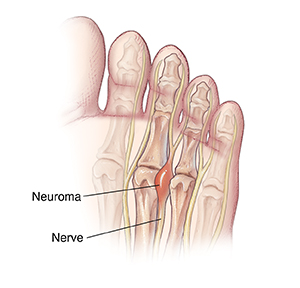
    • Neuroma formation
    • Entrapment of nerves in scar tissue.


    Underlying Disease Processes

    Key to managing symptoms of pain that have to do with a pre-existing condition like diabetes is to make sure that the condition is managed as well as possible.

    For example, if you have diabetes, keep your blood sugar under good control. If you have poor circulation, follow your physician’s instructions for diet, exercise and medications.

    Managing pre-existing conditions after surgery is as important as ever.


    Surgical Trauma

    Poor tissue coverage can be caused by the bone at the end of your residual limb not being properly trimmed at the time of surgery, and this can cause pain when you wear your prosthesis. If padding and other methods are not successful, surgery may be required to revise your residual limb so as to decrease your pain and allow you to wear your prosthesis.


    Neuromas

    A neuroma is a collection, or bundle, of nerve endings that forms under the skin of your residual limb. Think of it like a tangle of hair. It can become very sensitive, especially if the tangle is pressing against your prosthesis.

    Because neuromas are made up of nerve endings, possible treatments include medications that help with nerve pain, such as:

    • Non-steroidal anti-inflammatory medications
    • Specific antidepressants and anticonvulsants that have been found to be effective for
    • nerve pain
    • Steroid injections.

    Non-medication options include:

    • Ultrasound, which is essentially a machine that uses sound waves to generate heat within a body part. It can help increase blood flow so that inflammation and swelling can be decreased
    • Massage, which also helps decrease inflammation and also helps desensitize your residual limb to touch
    • Vibration, which creates a mild shaking to contract muscles, and to decrease inflammation and pain
    • Percussion/finger tapping at the point of pain, which also helps with desensitization of your residual limb
    • Acupuncture, manipulating thin, solid needles that have been inserted into specific pressure points in the skin, which has been helpful for some
    • TENS (transcutaneous electrical nerve stimulation), which produces a sensation of mild pins and needles, overriding some of the pain that your body is producing.

    Modifying the prosthesis socket to prevent rubbing at the sensitive part of your limb may also be helpful.

    Unfortunately, surgery to remove neuromas is not usually successful, because they often just reform.


    Heterotopic Bone

    Occasionally, excess bone forms abnormally around the end of the amputated limb; this is sometimes called a “bone spur.” The “extra” bone may cause pressure points that interfere with the fit of your prosthesis; this occurs more frequently in children than adults. If the problem cannot be solved with changes to the prosthesis, surgery can remove the excess bone. For children, this is best done after the bone stops growing.


    Entrapment of Nerves in Scar Tissue

    As your incision begins to heal, your doctor will let you know when you can start massaging your residual limb. This will help to prevent nerves from being “caught” in scar tissue. You will also be taught how to wrap your limb using elastic wraps. This not only helps to prevent scarring but also helps with prosthesis fit.


    Managing Pain

    No matter the cause of residual limb pain, there are some principles that can be followed to help you manage your pain.

    • Begin exercises as soon as your surgeon allows it. Standing, walking and muscle stretching not only improve your general health, but also interrupt pain signals.
    • Desensitize your residual limb, following the instructions of your physical or occupational therapist; this includes both massaging and wrapping your residual limb.
    • Keep a pain journal. Keeping track of your symptoms and sharing them with your healthcare team will help you find the right treatments for you.
    • Practice relaxation. We know that tension and stress increase pain. It is estimated that 50 percent of pain can be reduced by relaxation.


    About the Amputee Coalition

    The Amputee Coalition is a donor-supported, nonprofit voluntary health organization serving the over 5.6 million people with limb loss and limb difference in the United States.

    For more information, please call 888-267-5669 or visit amputee-coalition.org.


    Acknowledgements

    This project was supported, in part, by grant number 90LLRC0001-01-00, from the Administration for Community Living, U.S. Department of Health and Human Services, Washington, D.C. 20201. Grantees undertaking projects under government sponsorship are encouraged to express freely their findings and conclusions. Points of view or opinions do not, therefore, necessarily represent official Administration for Community Living policy.

    Source link

  • Little League Elbow | Nemours KidsHealth

    Little League Elbow | Nemours KidsHealth

    What Is Little League Elbow?

    Little League elbow is an overuse injury to the elbow caused by repetitive throwing. Most cases happen in pitchers, but any young athlete who throws a lot can get the condition.

    What Are the Signs & Symptoms of Little League Elbow?

    Kids with Little League elbow have pain on the inner part of their elbow. At first, the elbow may hurt only during or right after throwing. But without treatment, the elbow can start hurting all the time. The pain usually starts gradually, but can happen after one throw if the athlete has been making the same motions often.

    What Causes Little League Elbow?

    Little League elbow is an overuse injury (also called a repetitive stress injury). Overuse injuries happen because the same motion is repeated over and over again.

    In Little League elbow, repeated throwing injures a growth plate in the elbow. A growth plate is a layer of cartilage near the end of a bone where most of the bone’s growth happens. It’s weaker and more at risk for injury than the rest of the bone.

    Who Gets Little League Elbow?

    Little League elbow happens most often in pitchers. But anyone who throws a lot, including catchers, infielders, and outfielders, can get it.

    Most cases are in kids and teens 8–15 years old. They’re still growing, so their bones still have growth plates. Bones that are done growing don’t have growth plates. Elbow pain after this age likely is not Little League elbow.

    How Is Little League Elbow Diagnosed?

    Health care providers diagnose Little League elbow by: 

    • asking about sports and activities
    • doing an exam of the elbow, observing range of motion and doing strength tests
    • getting X-rays (X-rays can be normal in Little League elbow but can show other problems in the elbow)
    • comparing the affected elbow with the unaffected elbow

    How Is Little League Elbow Treated?

    Kids with Little League elbow must take a break from all throwing for about 6 weeks. For pain and swelling, they can:

    • Put ice or a cold pack on the elbow every 1–2 hours for 20 minutes at a time. Put a thin towel between the ice and the skin to protect it from the cold. 
    • Take ibuprofen (Advil, Motrin, or store brand) or naproxen (Aleve, Naprosyn, or store brand) if the health care provider says it’s OK. Follow the package directions for how much to give and how often to give it. Kids should always take the medicines with food.

    When pain and swelling ease, health care providers usually recommend physical therapy or another exercise program. Before returning to play, it might help some kids to work on how they throw with a pitching coach or physical therapist.

    When Can Kids With Little League Elbow Go Back to Throwing?

    After a rest period and physical therapy or another exercise program, athletes with Little League elbow can slowly return to pitching if they:

    • do not have any elbow pain
    • have full strength in their arm
    • can bend and straighten their elbow fully

    Athletes with Little League elbow need to work with their health care provider and coach to create a return to pitching program. The program should:

    • slowly increase the number, distance, and intensity of pitches over 6–8 weeks
    • say how many pitches can be thrown a day
    • say what distance it is OK to throw

    They shouldn’t go back to throwing until their health care provider says it’s OK. Going back too early can permanently damage the elbow.

    Can Little League Elbow Be Prevented?

    To help prevent Little League elbow, young athletes should:

    • Take a break from throwing for 3–6 months a year. They can play another sport that doesn’t involve throwing, like soccer or swimming.
    • Make sure they throw correctly.

    They also should follow pitching guidelines that specify:

    • how many pitches are OK for each age
    • what kind of pitches are OK for each age
    • how much rest is needed between practices and games

    You can find information online about pitching guidelines at:

    How Can Parents Help?

    To help kids with Little League elbow:

    • Make sure they follow the health care provider’s recommendations for rest and exercises.
    • Make sure they don’t go back to throwing until the doctor says it’s OK. Throwing too soon can cause permanent elbow damage.
    • When they’re back to throwing, be sure the pitching guidelines are followed. Someone from the team should keep track of pitches. If no one does, you may need to do it yourself.
    • Teach your child that if something hurts during training or a game, they should stop playing right away. If the pain continues, your child needs to get checked by a coach, trainer, and health care provider before returning to play.

    Source link

  • Understanding Parkinsonism | Parkinson’s Foundation

    Understanding Parkinsonism | Parkinson’s Foundation

    Parkinsonism: A set of movement symptoms associated with Parkinson’s disease (PD) and other disorders. It is important to understand that parkinsonism describes symptoms rather than a specific disorder.

    Symptoms include:

    • Slow Movements
    • Stiffness
    • Walking and balance problems
    • Tremor

    Parkinsonism is a name for a group of movement symptoms found in several conditions, including Parkinson’s disease. These symptoms include slow movements, stiffness, tremor and problems with walking and balance. Parkinsonism disorders can also involve non-movement symptoms, such as changes in mood and thinking, sleep problems and issues with automatic body functions, like blood pressure.

    Types of Parkinsonism

    Parkinson’s Disease

    • A progressive brain disorder with movement and non- movement symptoms
    • Symptoms worsen slowly over time
    • Movement symptoms tend to improve with levodopa

    Atypical Parkinsonism

    • A group of progressive brain disorders with some of the symptoms seen in PD
    • Symptoms are often more serious early on and progress quicker
    • Movement symptoms improve less or not at all with levodopa

    Secondary Parkinsonism

    • Conditions that mimic PD but are not always progressive
    • Symptoms may improve by treating the underlying cause
    • Movement symptoms usually do not improve with levodopa

    Atypical Parkinsonism Disorders

    Atypical parkinsonism disorders have specific traits that may help set them apart from each other.

    Dementia with Lewy Bodies (DLB)

    • Progressive short-term memory loss and confusion
    • Difficulty focusing or staying alert
    • Seeing things that are not there, like shadowy figures in your side vision

    Multiple System Atrophy (MSA)

    • Lightheadedness when standing up or wide swings in blood pressure
    • Difficulty controlling the bladder

    Progressive Supranuclear Palsy (PSP)

    • Slow or limited eye movements, both upward and downward
    • Serious balance problems and frequent falls early on
    • Early changes in thinking and behavior
    • Slurred or hoarse speech

    Corticobasal Degeneration (CBD)

    • Muscle tightness and jerking movements
    • Abnormal hand and feet postures (called dystonia)
    • Trouble with coordination

    Diagnosing Parkinsonism

    There is no single test for diagnosing parkinsonism disorders. Your neurologist will review your medical history, symptoms and medications, perform a neurological exam and may order brain scans (like an MRI or DaTscan) or a skin biopsy. How your symptoms respond to levodopa can also provide helpful clues.

    Since these disorders share similar symptoms, getting the right diagnosis can sometimes be tricky. It is important to work with a neurologist experienced in parkinsonism. Certain neurologists, known as movement disorders specialists, have extra training in this area. For help finding a specialist, contact our Helpline.

    Treating Atypical Parkinsonism

    Levodopa may not be effective for all types of atypical parkinsonism, but other medications and lifestyle changes may help reduce symptoms. Physical and occupational therapy, regular exercise and speech therapy are key to staying as active and independent as possible.

    Look for a neurologist who can help you build a care team tailored to your needs, including rehabilitation specialists and, if necessary, a social worker, psychiatrist, sleep doctor, urologist or other health professionals.

    Secondary Parkinsonism

    Some types of parkinsonism may improve if the underlying cause is treated. For example, drug-induced parkinsonism, a type of secondary parkinsonism, often improves after stopping the medication. Drugs commonly linked to this include antipsychotics, anti-nausea medications and some blood pressure drugs. Normal pressure hydrocephalus, which causes memory loss and walking issues, can be treated with a tube to drain excess fluid. Treatment for other forms of secondary parkinsonism depends on the specific cause.

    Tips for Navigating Parkinsonism

    • If unsure about your diagnosis, ask your doctor for clarification. Keep in mind that a specific diagnosis may take time.
    • Stay informed. Understanding your treatment options and available resources can help improve quality of life.
    • Connect with others with a similar diagnosis to share experiences, coping strategies and resources.
    • Focus on what you can control, like exercise, nutrition, sleep and staying engaged in activities and relationships that are important to you.

    Resources

    Source link

  • Driving | Parkinson’s Foundation

    Driving | Parkinson’s Foundation

    Smiling man in a suit driving a car

    Driving allows personal freedom, control and independence. Many people with Parkinson’s disease (PD) continue to drive safely long after their diagnosis.

    While Parkinson’s progression and medication side effects may affect a person’s driving ability, the diagnosis alone does not tell the whole story. The ability to drive with Parkinson’s depends on a person’s specific symptoms, as well as the presence of other age-related changes.

    Though Parkinson’s may present driving challenges, there are many ways to maintain independence. If you are facing driving challenges, the below information can help you take control of your transportation needs.

    How can Parkinson’s affect my driving?

    Driving is a complex skill. Parkinson’s-related physical, emotional and mental changes may affect your ability to drive safely.

    • Parkinson’s can cause your arms, hands or legs to shake even when you are relaxed. PD can also make it harder for you to keep your balance or start to move when you have been still.
    • You may not be able to:
      • React quickly to a road hazard.
      • Turn the steering wheel, push down on the gas pedal or brake as quickly as you may need or with precision.
    • Depth perception can affect your ability to change lanes, park, etc.
    • Changes to cognition may affect your ability to process all the complex actions that occur in order to drive safely.

    Many Parkinson’s medicines can also reduce your ability to drive safely. Common medications — including carbidopa/levodopa (Sinemet), amantadine, dopamine agonists and anticholinergics — may produce side effects such as sleepiness, dizziness, blurred vision and confusion. Not everyone experiences these side effects, and they may be decreased by simple medication adjustments.

    Can I still drive with PD?

    The ability to drive with Parkinson’s depends largely on your individual circumstances. Driving with PD tends to be safest in the early stages of the disease. Taking medications that control your symptoms can also improve your driving ability and staying active can help you keep the muscle strength you need to drive.

    How do I know if I can drive safely?

    It may be difficult to determine when, or if, you should limit your driving. Resources are available to help you better understand your own driving ability:

    Ask a trusted friend or family member for honest input about your driving skills.

    Take a driving assessment. Here’s how:

    • Assessments are available through your local DMV.
    • Your doctor also may recommend a Driving Rehabilitation Specialist’s (DRS) assessment. These professionals give on- and off-road tests to see if, and how, PD affects your driving. The specialist may offer driving skill improvement training if you can still drive safely. Call the Association for Driver Rehabilitation Specialists at 866-672-9466, check the directory for a DRS near you, or ask your doctor for a referral to a DRS.

    What can I do if I have to reduce my driving?

    If you must cut back on or give up your driving, you can still maintain your independence. Consult these driving alternatives to help identify which form of transportation best suits you:

    • Take public transportation such as a bus, subway or train. Bus passes are often offered for a reduced fee to the elderly and people living with disabilities. Call your local public transportation office to get information on discounts and find out what routes to take.
    • Take a taxi or car service, especially for quick or spontaneous errands. There are many apps you can download to your smartphone to request transportation.
    • Ask family and friends to drive you. One person may be willing to take you to the grocery store weekly and another might volunteer to pick up your medications from the pharmacy.
    • Ask about the ability of van service if you live in an independent or assisted living facility. Many facilities offer this service to take residents to appointments. Be sure to reserve this service in advance.
    • Check for alternative transportation options with your local city/town government and the local community center. Special shuttle or van services may be available for people with disabilities.
    • Contact your local religious organization to find out if they have a committee of volunteers who drive community members to different destinations.

    Who can help with transportation?

    Many services are available to help you locate transportation in your area. Consult these organizations to get started:

    Learn More

    Watch the DriveWise® program videos: Driving with Parkinson’s 1 and Driving with Parkinson’s 2, produced by Beth Israel Deaconess Medical Center, a Parkinson’s Foundation Center of Excellence. Ask about any similar programs at hospitals, driving schools, rehabilitation facilities and state motor vehicle departments in your area.

    Page reviewed by Dr. Bhavana Patel, Movement Disorders Neurologist at the University of Florida, a Parkinson’s Foundation Center of Excellence.

    Source link

  • Osteoporosis: What you need to know

    Osteoporosis: What you need to know

    Did you know that our bones are living tissue? To keep them strong, our bodies break down old bone and replace them with new bone. When we are young, we build more bone than we break down. But as we get older, bone can break down more often than it gets replaced. This can lead to osteoporosis.

    Osteoporosis can develop if your bone mass decreases or the structure of your bones changes. Osteoporosis makes your bones weak and more likely to fracture (break).

    Osteoporosis is called a “silent” disease because it doesn’t show any obvious symptoms. Many people do not know they have it until they break a bone after a minor injury or movement. Something as simple as falling from standing height, bending, lifting, or even intense coughing may cause a fracture. Osteoporosis can develop in any bone, but it’s most common in the bones of your hips, wrists, and vertebrae in the spine.


    Who is at risk for osteoporosis?

    While anyone can develop osteoporosis, the risk increases as you get older. Osteoporosis is more common in people with any of these risk factors:

    • Sex. Women are more likely to have osteoporosis than men, especially after menopause. White and Asian women are at the highest risk. African American and Hispanic women are at a slightly lower risk. While men are less likely to have osteoporosis, White men are at a higher risk than other groups of their sex.
    • Age. As you age, you lose more bone than you build. Women over age 65 are recommended to get screened for osteoporosis by their health provider. There are no official screening recommendations for men, but their risk increases after age 70.*
    • Family history. Having a parent with osteoporosis or hip fracture increases risk.
    • Body type. Being slim or thin-boned increases risk because there is less bone to lose.
    • Hormones. Hormone levels can affect risk for both men and women. Low estrogen levels in women due to menopause, hormone disorders, or extreme physical activity can weaken bones. Low levels of testosterone in men also raises their risk for osteoporosis.
    • Nutrition. A diet low in calcium, vitamin D, or protein can raise risk.
    • Lifestyle factors. These include heavy alcohol use, smoking, not being physically active, or being on bedrest for extended periods of time can all raise risk.
    • Medications. Long-term use of some medications may increase risk. These include corticosteroids, proton pump inhibitors, and medicines to treat epilepsy. Some cancer medications and glucocorticoid steroids can also increase risk.
    • Medical conditions. Some medical conditions increase risk, including:

    How is osteoporosis diagnosed?

    A health care provider can usually diagnose osteoporosis with a routine screening. They may ask you about your medical and family history and whether you have ever broken a bone. They may also want to know if you have lost height or weight or if you are losing muscle strength. Talk to your doctor if your posture, balance, and gait (how you walk) have changed.

    The most common test for osteoporosis is a bone mineral density test, which measures how strong your bones are and predicts your risk of bone fractures.

    How is osteoporosis treated?

    If you have the disease, your doctor will likely recommend treatment designed to slow or stop bone loss and help prevent fractures. They may recommend eating a balanced diet with enough calcium, vitamin D, and protein. They may also recommend other lifestyle changes or prescribe medicine to slow bone loss or rebuild bone. Medications can cause side effects, so talk to your doctor or pharmacist if you have questions about what you’re taking.

    Regular physical activity is important if you have osteoporosis, but certain activities may put a strain on high-risk parts of the body. Avoid high-impact exercise and movements that twist your spine (like swinging a golf club) or heavily bend the waist (like sit-ups and toe touches). A physical therapist, rehabilitation medicine specialist, or physiologist can help you make a safe exercise program. Caregivers can also help someone with osteoporosis exercise in a safe way.

    Can it be prevented?

    Keeping bones strong throughout your life can help lower your risk for osteoporosis, so eat a healthy diet rich in calcium and vitamin D, get regular physical activity, limit alcohol intake, and don’t smoke. It’s also important to have regular checkups and tell your doctor if you’ve had a fall or broken a bone.

    Living with osteoporosis

    Whether you have osteoporosis or are trying to prevent it, proper nutrition and certain exercises are good for bone health. Strong muscles help you balance and reduce your risk of falling or breaking bones. People tend to lose muscle as they age, but a combination of weight-bearing exercises, resistance training, and balance training can help. Talk to your health care provider about what’s best for you before starting any exercise program.

    The National Institute of Arthritis and Musculoskeletal and Skin Diseases also has information on exercising for bone health.

    If you have osteoporosis, talk with others who have it and reach out to family and friends for support. Learning about the disease can equip you to participate in decisions about your care and help preserve your mobility and independence!

    *Editor’s note: As of this publication, the U.S. Preventive Services Task Force’s osteoporosis screening recommendations are under review. Learn more about the review process and updates here.

    Source link

  • ClinicalTrials.gov: Menorrhagia

    ClinicalTrials.gov: Menorrhagia

    Source: National Institutes of Health – From the National Institutes of Health
    Related MedlinePlus Pages: Menstruation

    Source link

  • Lower Your COPD Risk at Any Age

    Lower Your COPD Risk at Any Age

    Source: National Heart, Lung, and Blood Institute – From the National Institutes of Health Video
    Related MedlinePlus Pages: COPD

    Source link

  • ClinicalTrials.gov: Osteomalacia

    ClinicalTrials.gov: Osteomalacia

    Source: National Institutes of Health – From the National Institutes of Health
    Related MedlinePlus Pages: Vitamin D Deficiency

    Source link