

The name sounds like something from a children’s book. Pott’s puffy tumor is not a tumor at all, and the thing it actually describes is far less charming: the frontal bone of the skull being destroyed by infection, with a pocket of pus collecting between the bone and the tissue covering it.
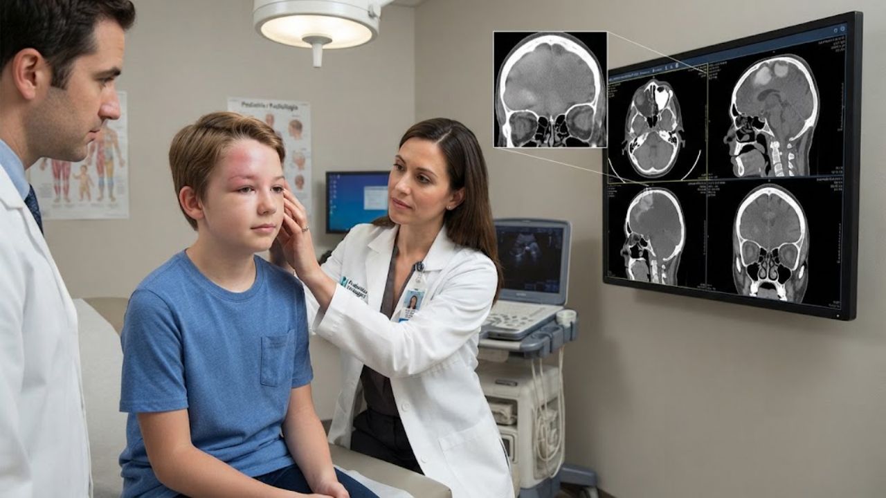
A 13-year-old boy in Pakistan arrived at hospital having fallen 15 days earlier. He now had a headache, vomiting, and two days of swelling around his left eye and left forehead, with reddened skin and tenderness over the frontal sinus. The clinical question put to radiology was straightforward: rule out a fracture and a subdural hematoma.
The CT found neither. The case was published in June in Cureus by Muniba Manzoor of Combined Military Hospital in Rawalpindi and colleagues at hospitals in Islamabad and Dublin.
The Trauma Story Was a Coincidence
His neurological examination was normal, with intact cranial nerves, no nystagmus and no ophthalmoplegia. Eye movements were full and painless. Almost all laboratory values came back normal, including C-reactive protein at 3.3 mg/L against a threshold of 5 and a neutrophil percentage of 52 within a range of 40 to 70. His white blood cell count was the single abnormal result, at 11,500 cells per microliter against an upper limit of 11,000.
Non-contrast CT showed both frontal sinuses opacified and filled with a soft tissue collection. In the bone window, the left frontal bone showed focal erosion with irregular destruction and marked thinning of both the inner and outer tables. Cortical margins were disrupted, bone density was reduced and the normal trabecular architecture had been lost. Those are the radiological hallmarks of osteomyelitis.
Further images showed a hypodense collection in the soft tissue of the forehead, an epidural abscess in the left frontal region, and a subperiosteal abscess. A sagittal view revealed a hypodense area in the adjacent frontal lobe itself, raising concern for early spread into the brain.
The fall had brought him in. The disease had been building in his sinuses.
A Diagnosis from 1768 That Never Went Away
Percivall Pott, the London surgeon, first described the condition in 1768, initially in the context of head trauma. Untreated or inadequately treated frontal sinusitis is now the dominant cause. The word tumor in the name carries its older sense of a swelling rather than a growth.
The pathway is anatomical. Infection in the mucosa of the frontal sinus spreads into the diploic veins, the channels running through the spongy layer between the skull’s inner and outer tables. Those veins carry it into the bone, producing osteomyelitis, and pus then collects under the periosteum to form the abscess that gives the forehead its characteristic puffy swelling.
Children and adolescents are the classic population, and there is a developmental reason. Diploic vein vascularity peaks during adolescence while the frontal sinuses are still reaching adult size, which the authors say increases the risk of infection reaching surrounding bone. Less common triggers include intranasal drug use.
Broad-spectrum antibiotics made the condition rare, but did not eliminate it. A BMC Pediatrics report describes a 9-year-old with an epidural empyema on MRI and CT evidence of bone erosion whose blood cultures grew Streptococcus anginosus, and a pediatric case published last year documents an 8-year-old whose infection had already reached inside the skull.
What Happens If Nobody Catches It
The reason clinicians care about a condition this uncommon is the neighborhood. The infected bone sits directly against the front of the brain.
The recognized complications are epidural abscess, subdural empyema, cerebral abscess, meningitis, and cavernous sinus thrombosis. This patient already had an epidural abscess and a suspicious frontal lobe finding at the time of his first scan, roughly two weeks after the fall that prompted it.
The symptoms that should raise suspicion are ordinary enough to be missed: forehead swelling, fever, headache, nasal discharge, and swelling or redness around the eye. Vomiting, nausea, and visual or neurological changes appear as things progress. The condition is worth considering in any child with frontal sinusitis that is not responding adequately to antibiotics.
CT is generally the first-line imaging test because it shows bone erosion, sinus opacification, and subperiosteal collections clearly. MRI is added when intracranial complications are suspected, since it gives better soft-tissue detail and can characterize collections inside and outside the skull.
Ten Weeks of Antibiotics and No Operation
Standard management combines prolonged intravenous antibiotics with surgery, usually drainage of the abscess and debridement of dead bone, sometimes via endoscopic sinus surgery. An international consensus statement built on a Delphi process attempted to standardize an approach that has long varied between centers.
This boy did not have an operation. He was treated with moxifloxacin 400 mg followed by linezolid 400 mg, given for up to 10 weeks. The abscess resolved, he improved clinically, and the authors report complete recovery with no sign of recurrence.
What the authors frame as individualized is the choice of drug, not the absence of surgery. Other teams have used cefepime, metronidazole, and vancomycin. Antibiotic choice, they write, should follow local guidelines and resistance patterns, cover anaerobes and Gram-positive organisms, and cross the blood-brain barrier when there is intracranial involvement.
One inconsistency is worth noting for readers comparing details. The discussion section states that laboratory tests in this case were normal, while the case description and the published table record an elevated white cell count. The imaging findings, not the bloodwork, drove the diagnosis either way.
A single case cannot establish how often antibiotics alone will suffice. What it does illustrate is that a scan ordered for one reason found something considerably more urgent, and that a swollen forehead in a child deserves a real explanation. Parents should seek medical evaluation for forehead swelling accompanied by fever, headache, or eye symptoms rather than attributing it to a recent bump.
Key Questions Answered
Is Pott’s puffy tumor actually a tumor?
No. The name uses an older sense of tumor, meaning a swelling. It describes osteomyelitis of the frontal bone with a subperiosteal abscess, both caused by infection.
What causes it?
Most often, untreated or inadequately treated frontal sinusitis. Trauma is the other recognized cause, and it was the original context in which Percivall Pott described it.
How does a sinus infection reach the skull bone?
Through the diploic veins, which run within the spongy layer of the skull and carry infection from the sinus mucosa into the bone.
Why is it dangerous?
The infected bone sits directly against the brain. Recognized complications include epidural abscess, subdural empyema, brain abscess, meningitis and cavernous sinus thrombosis.
How was this patient treated?
With intravenous moxifloxacin followed by linezolid for up to 10 weeks, without surgery. He recovered fully. Most cases combine antibiotics with surgical drainage.
What symptoms should prompt evaluation?
Forehead swelling with fever, headache, nasal discharge, or swelling and redness around the eye, particularly in a child whose sinusitis is not improving on antibiotics.


